Correctly Label the Following Muscles of the Neck.
Sign up for an account today. Replace figure with one that includes all muscles from table for example figure 107 from Marieb or 98 from Amerman.

Neck Muscle Anatomy Health Medicine And Anatomy Reference Pictures Anatomiya Cheloveka Anatomiya Myshcy
These muscles have two origins one on the sternum and the other on the clavicle.
. Interscapular- between shoulder blades. Muscles of the neck Musculi cervicales The muscles of the neck are muscles that cover the area of the neckThese muscles are mainly responsible for the movement of the head in all directions. Correctly label the muscles of the thoracic cavity and the abdomen.
Click on the tags below to find other quizzes on the same subject. They insert on the mastoid process of the temporal bone. They can flex or extend the head or can rotate the towards the shoulders.
Splenius capitis and splenius cervicis are a pair of superficial muscles in the back. Correctly label the following muscles of the anterior view. View F0F6E090-C906-4744-9569-76A37672BAC3jpeg from MAT MISC at Bunker Hill Community College.
Nuchal- back of neck. Muscle fiber sarcolemma mitochondria myofibril i band sarcoplasm sarcoplasmic reticulum z disc a band myofilaments noonallig 603 ar correctly label the following parts of a skeletal muscle fiber. This quiz has tags.
Rectus capitis posterior major and Rectus capitis posterior minor attach the inferior nuchal line of the occiput to the C2 and C1 vertebrae respectively. Located underneath the platysma on the sides of the neck are the sternocleidomastoid muscles. Depressor labii Frontal belly.
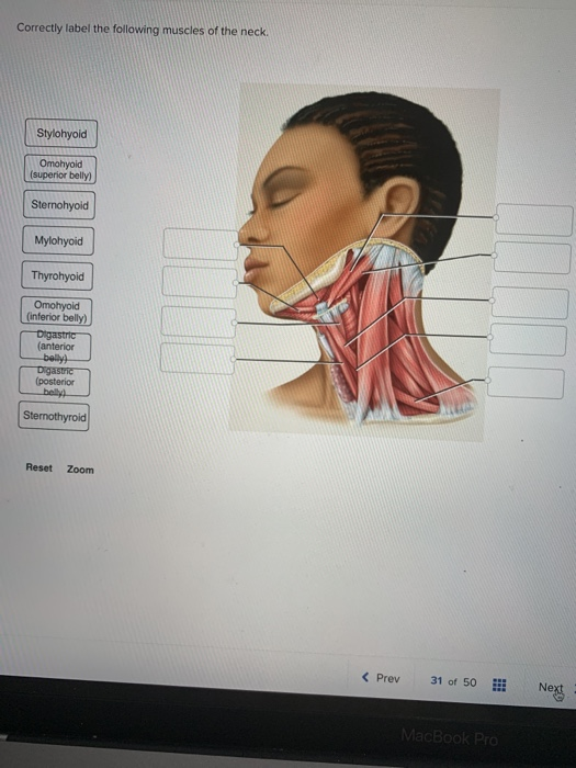
View Homework Help - 54592789-E9BC-4A06-96A2-E05E2C81BB6Bjpeg from BIO 203 at Bunker Hill Community College. Anatomy and Physiology questions and answers. Correctly label the following muscles of the neck.
Correctly label the following parts of a skeletal muscle fiber. Show transcribed image text Expert Answer. - Osmosis is an efficient enjoyable and social way to learn.
Correctly label the following muscles of facial expression. Correctly label the following anatomical features of the heart and thoracic cage correctly label the following vessels leading from and toward the anterior heart correctly label the following external anatomy of the posterior heart. Obliquus capitis superior also extends from the occiput to C1 while obliquus capitis inferior originates.
Experts are tested by Chegg as specialists in their subject area. Correctly label the muscles of the leg. On the flanks of the body medial to the rectus femoris the abdominal wall is composed of three layers.
Which term refers to a muscle that prevents a bone from moving during an action. Anatomy of the lymphatics of the neck Videos Flashcards High Yield Notes Practice Questions. Correctly label the following muscles of the neck.
Which of the following is a functional requirement of cardiac muscle tissue. Who are the experts. Stylohyoid Omohyoid superior belly Sternohyoid Mylohyoid Thyrohyoid inferior anterior belly posterior Sternothyroid Reset Zoom くPrev 31of 50Hİ Next.
Correctly label the following regions of the external anatomy. The occipitofrontalis muscle elevates the scalp and eyebrows. This is an online quiz called Anterior Neck Muscles.
Anterior lateral and posterior groups based on their position in the neckThe musculature of the neck is further divided into. Dont study it Osmose it. View Homework Help - AHCDW6SOL21pdf from AHCD 1011 at The University of Sydney.
Openings into transverse tubules sarcoplasmic reticulum sarcolemma mitochondria nucleus triad. We review their content and use your feedback to. One on each side of the neck.
There is a printable worksheet available for download here so you can take the quiz with pen and paper. What is the name of muscle A. 8 Correctly label the following muscles of the posterior view.
Correctly label the following regions of the external anatomy. Correctly label the following muscles of facial expression. The action for the vastus lateralis muscle is to _____ the knee.
Learn and reinforce your understanding of Anatomy of the lymphatics of the neck. Correctly label the following muscles of the neck. Correctly label the following muscles of facial expression.
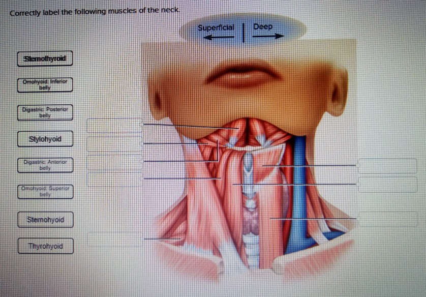
Place the following terms in order moving from superficial to deep. Also Know what muscles are involved in neck extension. Under the platysma are two sternocleidomastoid muscles.
With one on each side of the neck these help flex the neck and rotate the head upward and side to side. Dorsum of hand- top of hand. They consist of 3 main groups of muscles.
The epicranius muscle is also very broad and covers most of the top of. Correctly label the following muscles of the neck. This is a back view of the right leg.
The muscle has a frontal belly and an occipital belly near the. Correctly label the following muscles of the neck SuperficialDeep Slemothyrod Omohyoid bely Digastnc. The muscles of the neck are present in four main groups.
Adjust credit for all students. View Correctly label the following muscles of the posterior viewpng from NURS MISC at University Of Connecticut. The suboccipital muscles act to rotate the head and extend the neck.
Correctly label the following muscles of the anterior. The orbicularis oris is a circular muscle that moves the lips and the orbicularis oculi is a circular muscle that closes the eye. Are these muscles labeled correclty.
Solved Correctly Label The Following Muscles Of The Neck Chegg Com

Human Muscle System Functions Diagram Facts Britannica
Solved Correctly Label The Following Muscles Of The Neck Chegg Com
0 Response to "Correctly Label the Following Muscles of the Neck."
Post a Comment